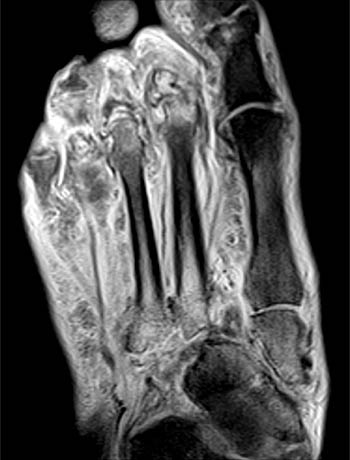
Foot Muscles Mri : Soleus Muscle Radiology Reference Article Radiopaedia Org : Mri of the soft tissues of the foot visualizes the fat cushions of the sole, heels, fingers and can show swelling, foci of infiltration and inflammation.
byAdmin•
0
Foot Muscles Mri : Soleus Muscle Radiology Reference Article Radiopaedia Org : Mri of the soft tissues of the foot visualizes the fat cushions of the sole, heels, fingers and can show swelling, foci of infiltration and inflammation.. Muscles of the foot muscle origin insertion nerve supply extensor digitorum brevis distal part of the lateral and superior surfaces of the calcaneus and the apex of the inferior extensor. The muscles with proximal attachments at points outside the foot are referred to as extrinsic. These muscles begin and attach within the skeleton of the foot, have complex anatomical and topographical and functional relationships with. Mri with hardware in foot? Lateral and medial processes of calcaneal tuberosity.
Related posts of foot muscle anatomy mri. Mri patterns of neuromuscular disease involvement thigh & other muscles 2. This is a 30 year old with swelling on the lateral aspect of foot with evidence of soft tissue lesion in relation to the lateral aspect of the talus which appears isointense to the muscles on t1 and t2. Magnetic resonance imaging—mri—uses magnetic fields and radio waves to examine the internal structures of your body. Metabolic and anatomic abnormalities identified, were grouped into muscular, neurovascular, and skin lesions.
Mri Of The Diabetic Foot Radsource from radsource.us Human anatomy for muscle, reproductive, and skeleton. Related posts of foot muscle anatomy mri. Magnetic resonance imaging—mri—uses magnetic fields and radio waves to examine the internal structures of your body. Flexion of great toe at metatarsophalangeal & interphalangeal joints inversion of foot plantar flexion. ► shoulder ► elbow ► wrist ► finger ► thumb. Thank you for your attention. Muscles of the foot are located on its rear and on the sole. Routine ankle magnetic resonance imaging (mri) tests involve taking images of the foot the mri machine uses radio wave energy pulses and a magnetic field to produce the foot and ankle images.
Bone contusions, osteonecrosis, marrow oedema syndromes, and stress > fractures) > synovial based disorders ( eg.
Indications for foot mri scan. Posted by radiologyer at 8:12 am. Gray's anatomy for students, 2nd ed. Related posts of foot muscle anatomy mri. Mri with hardware in foot? Resulting pet/mri images were reviewed by two radiologists. Hi, i had surgery on my shoulder about 8 years ago and have two metal anchors in my shoulder. The flexor digiti minimi brevis (flexor brevis minimi digiti, flexor digiti quinti brevis) lies under the metatarsal bone on the little toe, and resembles one of the interossei. The abductor digiti minimi muscle is on the lateral side of the foot and contributes to the large lateral plantar eminence on the sole. Muscles of the ankle and foot. Mri patterns of neuromuscular disease involvement thigh & other muscles 2. Learn about foot and ankle mri here. ► shoulder ► elbow ► wrist ► finger ► thumb.
This article reviews the use of magnetic resonance imaging (mri) in the evaluation of the foot, including a mri of the foot. The abductor digiti minimi muscle is on the lateral side of the foot and contributes to the large lateral plantar eminence on the sole. Foot positioned for axial images of the ankles; An overview of the intrinsic muscles of the foot including their origin, insertion, blood supply, innervation, function and clinical relevance. Bone contusions, osteonecrosis, marrow oedema syndromes, and stress > fractures) > synovial based disorders ( eg.
Mri Of The Ankle Detailed Anatomy W Radiology from w-radiology.com A magnetic resonance imaging (mri) was performed on a normal subject; The extrinsic muscles are located in the anterior and lateral compartments of the leg. Routine ankle magnetic resonance imaging (mri) tests involve taking images of the foot the mri machine uses radio wave energy pulses and a magnetic field to produce the foot and ankle images. An overview of the intrinsic muscles of the foot including their origin, insertion, blood supply, innervation, function and clinical relevance. Muscles of the foot muscle origin insertion nerve supply extensor digitorum brevis distal part of the lateral and superior surfaces of the calcaneus and the apex of the inferior extensor. Metabolic and anatomic abnormalities identified, were grouped into muscular, neurovascular, and skin lesions. The flexor digiti minimi brevis (flexor brevis minimi digiti, flexor digiti quinti brevis) lies under the metatarsal bone on the little toe, and resembles one of the interossei. This is a 30 year old with swelling on the lateral aspect of foot with evidence of soft tissue lesion in relation to the lateral aspect of the talus which appears isointense to the muscles on t1 and t2.
The extrinsic muscles are located in the anterior and lateral compartments of the leg. Mri with hardware in foot? Lateral and medial processes of calcaneal tuberosity. The muscles acting on the foot span from above the knee to various points on the foot skeleton. Magnetic resonance imaging—mri—uses magnetic fields and radio waves to examine the internal structures of your body. Posted by radiologyer at 8:12 am. Thank you for your attention. Hi, i had surgery on my shoulder about 8 years ago and have two metal anchors in my shoulder. Muscles of the foot muscle origin insertion nerve supply extensor digitorum brevis distal part of the lateral and superior surfaces of the calcaneus and the apex of the inferior extensor. Gray's anatomy for students, 2nd ed. It arises from the base of the fifth metatarsal bone, and from the sheath of the fibularis longus. Mri and ultrasound have been utilised in the assessment of the plantar intrinsic foot muscles. Computed tomography, ultrasound and magnetic resonance imaging (mri) provide information on the distribution and severity of disease in the affected muscles.
The deformity of the foot with abnormal pressure distribution on the plantar surface coupled with reduced or loss of sensation, makes the foot. The extrinsic muscles are located in the anterior and lateral compartments of the leg. Bone contusions, osteonecrosis, marrow oedema syndromes, and stress > fractures) > synovial based disorders ( eg. Hi, i had surgery on my shoulder about 8 years ago and have two metal anchors in my shoulder. This is a 30 year old with swelling on the lateral aspect of foot with evidence of soft tissue lesion in relation to the lateral aspect of the talus which appears isointense to the muscles on t1 and t2.
Mri Of The Ankle Detailed Anatomy W Radiology from w-radiology.com The muscles acting on the foot span from above the knee to various points on the foot skeleton. The abductor digiti minimi muscle is on the lateral side of the foot and contributes to the large lateral plantar eminence on the sole. Resulting pet/mri images were reviewed by two radiologists. Foot positioned for axial images of the ankles; Gray's anatomy for students, 2nd ed. ► hip ► pelvis ► thigh ► knee ► lower extremity/shin ► ankle ► foot. ► shoulder ► elbow ► wrist ► finger ► thumb. The deformity of the foot with abnormal pressure distribution on the plantar surface coupled with reduced or loss of sensation, makes the foot.
Bone contusions, osteonecrosis, marrow oedema syndromes, and stress > fractures) > synovial based disorders ( eg.
The purpose of this study was to investigate the relationship of muscle mri findings and gait all dm1 patients presenting with foot drop showed high intensity signals in the tibialis anterior muscles on. Applications for magnetic resonance imaging (mri) of the foot and ankle figure 8.4 image planes for foot and ankle mri. Gray's anatomy for students, 2nd ed. Learn about foot and ankle mri here. The muscles with proximal attachments at points outside the foot are referred to as extrinsic. Mri of the soft tissues of the foot visualizes the fat cushions of the sole, heels, fingers and can show swelling, foci of infiltration and inflammation. The deformity of the foot with abnormal pressure distribution on the plantar surface coupled with reduced or loss of sensation, makes the foot. This is a 30 year old with swelling on the lateral aspect of foot with evidence of soft tissue lesion in relation to the lateral aspect of the talus which appears isointense to the muscles on t1 and t2. Resulting pet/mri images were reviewed by two radiologists. Mri with hardware in foot? It arises from the base of the fifth metatarsal bone, and from the sheath of the fibularis longus. ► shoulder ► elbow ► wrist ► finger ► thumb. Thank you for your attention.